The goal of the core services is to accelerate the development of pediatric kidney atlas by providing kidney tissue for omics studies and iPSC lines for building kidney and molecular studies. The core infrastructure is partly supported by the Kidney Translational Research Center (KTRC) and iPSC line distribution is jointly collaborated with the ATLAS-D2K data center in the (Re)Building a Kidney (RBK) and GenitoUrinary Development Molecular Anatomy Project (GuDMAP) consortia.
The PCEN biological cores support innovation in modeling kidney development and disease using iPSC cell lines and multiomic single cell and spatial biology studies. The areas supported by the PCEN include kidney development, pediatric kidney diseases (CAKUT, glomerular diseases), tissue engineering, disease modeling, tissue-based research, multiomics studies related to single cell and spatially resolved methods to understand kidney biology.
Human iPSC and Organoid Core
The induced pluripotent stem cells (iPSC) core has set up an infrastructure of inter-institutional regulatory approvals to provide a number of human iPSC cell lines including parent and lineage and cell type specific reporter lines that can be differentiated into various kidney cell types. These cell lines are distributed throughout the world for various aspects of kidney development, disease modelling and tissue engineering.
Pediatric Kidney Tissue Core
Pediatric kidney tissue will be preserved in different media for broad application in single cell omics technologies. Planned preservations include FFPE blocks, Fresh frozen OCT-embedded blocks, flash frozen tissue, cryoprotected fixed frozen OCT-embedded tissue blocks and parent and reporter human iPSC lines. The source of pediatric deceased donor kidney tissue is from a network of organ procurement centers coordinated by Gloria Pryhuber, MD at University of Rochester Medical Center and nephrectomy and biopsy cases at Washington University coordinated by Sanjay Jain, MD, PhD. As the repository is being built, we anticipate pediatric kidney tissue to be available upon request and necessary regulatory approvals in 2025.
Single Cell & Spatial Biology Core
Accelerating pediatric kidney disease research
This core is directed by Drs. Sanjay Jain and Michael Eadon. The PCEN investigators are leaders in the field of single cell and spatial biology with expertise ranging from tissue acquisition, processing, data generation and analytics. They have generated a wealth of multiomics data from human kidneys. The services here will provide resources to accelerate pediatric kidney disease research by providing focused support for cell type and spatial expression queries, consultation in designing spatial transcriptomics experiments and potentially wet lab support for conducting spatial biology experiments.
Core Director

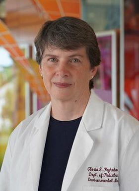
Gloria Pryhuber
Professor of Pediatrics, Neonatology
University of Rochester Medical Center, Rochester, New York
Gloria Pryhuber is a physician-scientist, currently Professor of Pediatrics (Neonatology) and Environmental Medicine at the University of Rochester Trained at the University for Cincinnati, Cincinnati Children’s Hospital Medical Center Pulmonary Biology Division. She has actively studied pathogenesis of chronic lung disease, especially following premature birth, for more than 25 years. In addition to leadership in clinical-translational studies, she is the PI for Phase I and Phase II of the Lung Development Molecular Atlas Program Human Tissue Core (LungMAP HTC) and now the HuBMAP-Lung Tissue Mapping Center. In partnership with the UNOS national transplant network, using low post-mortem interval, transplant organ recovery protocols, she created and manages the unique BioRepository for Investigation of Neonatal Diseases of the Lung (BRINDL), containing consented, transplant-quality, biospecimens of pediatric research tissues including trachea and lung tissue, peripheral blood mononuclear cells (PBMC), lymph node, spleen and thymus, and has provided over 40 research laboratories with unique opportunities to explore the developing human respiratory tract and immune system in an unusually holistic manner. HTC BRINDL samples have been used and published, by Dr. Pryhuber’s laboratory and by collaborators, for flow and mass cytometry, single cell and nuclear transcriptomics, epigenomics, unbiased mass spectrometry lipid and protein identification, highly multiplexed immunofluorescence and spatial transcriptomics. Most recently, in collaboration with Dr. Jain, she brings this infrastructure and capability to the PCEN pKidBIO Biospecimens Core for biobanking of pediatric human kidney, ureter and bladder, enthusiastic to support an outstanding team of scientists, students and clinical subspecialists in urology and nephrology.
Core Associate Director

Sanjay Jain, MD, PhD
Professor of Medicine, Nephrology
Professor of Pediatrics, Molecular Genetics and Genomics Program
Director Kidney Translational Research Center
Washington University in St. Louis School of Medicine
Sanjay Jain is a Professor of Medicine, Pediatrics and Pathology & Immunology at the Washington University School of Medicine in St. Louis, Missouri, USA (WUSM). His laboratory focuses on how kidneys and the lower urinary tract develop and organize to maintain homeostasis across lifespan in health and disease. His has defined key developmental pathways and mechanisms that regulate the joining of primitive ureter and bladder, initiation of the collecting system and branching morphogenesis of the kidney and genetic mutations associated with CAKUT. He leads multiple NIH-sponsored atlas efforts to map healthy and disease states in the human kidney including HuBMAP, KPMP, RBK/GUDMAP and Pediatric Center of Excellence in Nephrology. The team has identified, validated and mapped ~100 cell identities in the kidney including healthy and injured cells and defined genes and pathways that help recovery or predict decline in kidney function.
Core Co-Investigator

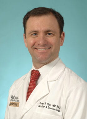
Joseph P. Gaut, MD, PhD
Ladenson Professor of Pathology, Pathology and Immunology
Division Chief, Anatomic and Molecular Pathology
Washington University in St. Louis School of Medicine
Joseph Gaut, MD, PhD is the Ladenson Professor of Pathology and Immunology, the Division Chief of Anatomic and Molecular Pathology, and the Medical Director of the Barnes-Jewish Hospital Histology Laboratory. Gaut received his MD and PhD degrees from Washington University School of Medicine. He completed an internship in General Surgery at Barnes Hospital/Washington University and a post-doctoral fellowship at Washington University with Jack Ladenson, PhD. He completed his Anatomic Pathology residency at Massachusetts General Hospital, Clinical Pathology residency at Washington University, and fellowships in renal and surgical pathology at Washington University. Gaut’s clinical focus is in the area of renal pathology. His research is focused on improved diagnostic methods for renal disease. He has published extensively in genetic glomerular disease, acute kidney injury (AKI) pathology, and emerging AKI biomarkers. More recently, Gaut has investigated image analysis methods for evaluation of organ quality prior to transplantation. He has collaborated on several consortia projects including the Kidney Precision Medicine Project and the Human Biomolecular Atlas Program.